Skin Lesions Write for Us
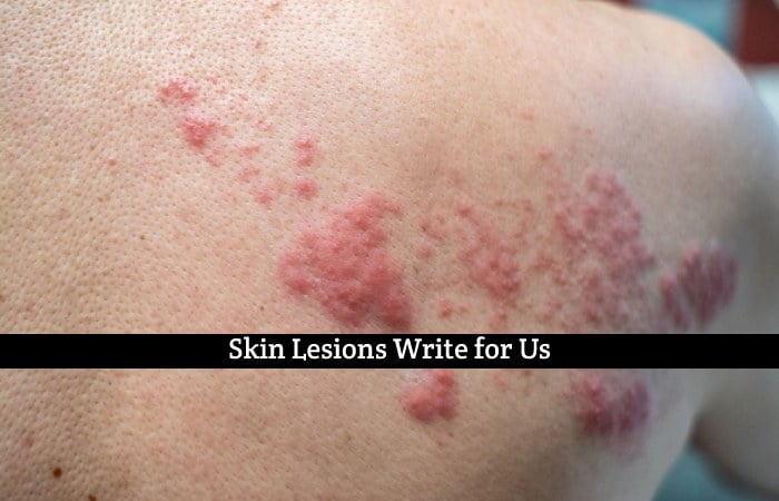
Skin lesions are abnormal areas of the skin that differ in color, texture, or appearance from the surrounding skin. Various factors, including infections, injuries, allergies, and underlying medical conditions, can cause them. Skin lesions can present as bumps, lumps, sores, rashes, or discolorations and may be benign or malignant.
Common skin lesions include moles, warts, acne, eczema, psoriasis, and skin cancer. Identifying and diagnosing skin lesions is crucial as they can range from harmless to potentially life-threatening. Medical professionals, such as dermatologists, use visual inspection, medical history, and sometimes biopsies to determine the nature of the lesion and recommend appropriate treatment.
It is essential to monitor any changes in existing skin lesions or the arrival of new ones, as early detection and timely medical intervention can significantly impact the outcome and overall skin health. Regular skin checks and sun protection are essential for maintaining healthy skin & reducing the risk of harmful skin lesions.
Note:- Before submitting articles, please read our guest writing policies.
Skin Lesions Write for Us Submissions: contact@healthsunlimited.com
What is a skin Lesions?
A skin lesion is an abnormal area or change on the skin’s surface, which may differ in color, texture, size, or shape from the surrounding skin. Skin lesions can result from various causes, including infections, injuries, inflammatory conditions, allergies, and underlying medical conditions. They can appear as localized areas of discoloration, bumps, lumps, blisters, sores, or ulcers.
There are two critical categories of skin lesions: primary & secondary.
Primary Skin Lesions:
- Macules: Small, flat, and colored spots, such as freckles or petechiae (tiny red or purple dots).
- So, Papules: Small, raised, solid bumps, like pimples or insect bites.
- Nodules: Larger, firm, elevated lesions often extend into the skin’s deeper layers.
- Plaques: Flat, raised, and often scaly patches commonly seen in conditions like psoriasis.
- Vesicles: Small fluid-filled blisters, like those seen in herpes or contact dermatitis.
- Bullae: Larger fluid-filled blisters, as seen in some types of dermatitis.
- So, Pustules: Pus-filled lesions, like acne or bacterial skin infections.
Secondary Skin Lesions:
- Crusts: Dried serum, blood, or pus overlying an ulcer or erosion.
- Scales: Flaking, dry skin, as seen in psoriasis or seborrheic dermatitis.
- Declines: Superficial loss of the epidermis, often resulting from a ruptured blister or scratch.
- Ulcers: The deeper epidermis and dermis loss create an open sore.
- Fissures: Cracks or splits in the skin, commonly seen in dry and thickened areas.
- Atrophy: Thinning or depression of the skin surface, as seen in some scars.
Skin lesions can be harmless and temporary, such as those resulting from insect bites or minor irritations. However, they can also indicate more severe conditions, including skin cancer. It is essential to monitor any changes in existing lesions or the arrival of new ones and seek medical evaluation if there are concerns about the lesion’s nature or if it exhibits concerning features (e.g., asymmetry, irregular borders, changes in color or size).
Dermatologists and other healthcare professionals are trained to evaluate and diagnose skin lesions through visual inspection, patient history, and, if necessary, by performing a skin biopsy for laboratory examination. Early detection and proper management of skin lesions are crucial to ensure timely and appropriate treatment and to preserve skin health. Additionally, practicing sun protection measures, regular skin checks, and seeking medical attention for any concerning lesions can help reduce the risk of possibly harmful skin conditions.
How do you Document a Skin Lesion?
Documenting a skin lesion involves several key steps:
- Description: Note the lesion’s size, shape, color, texture, and associated symptoms like pain, itching, or tenderness.
- Location: Record the precise location of the lesion on the body using anatomical landmarks for reference.
- Photograph: Take clear pictures from different angles and distances to document the lesion’s appearance for future reference and comparison.
- Patient Information: Include relevant patient details, such as name, age, medical history, and other relevant information.
- Date and Time: Document the date and time of the assessment or examination.
- Diagnosis: If possible, include the initial clinical impression or diagnosis of the skin lesion.
- Follow-up: If needed, schedule follow-up assessments to monitor changes or progression of the lesion.
Accurate documentation aids healthcare professionals in tracking the lesion’s evolution, assisting in diagnosis, and determining appropriate treatment plans.
We accept guest posts on Below Topics.
- Health
- Beauty
- Fashion
- Skin
- Hair
- Diet
- Nutritions
How to Submit Your Articles?
Before creating anything for our website, we ask that you carefully read our standards. Once your Post complies with our requirements, you can email it to us at contact@healthsunlimited.com
Why Write for Healths Unlimited – Skin Lesions Write for Us.

Search Terms Related to Skin Lesions Write for Us.
medical condition
integumentary system
organ system
body
skin
nails
glands
trauma
disease
Ghon lesions
varicella zoster virus
chickenpox
dental caries
Search Terms for Skin Lesions Write for Us.
Skin Lesions to submit an article
guest posting guidelines
become a guest blogger
become an author
Skin Lesions Submits Post
guest posts wanted
suggest a post
Skin Lesions guest post
Skin Lesions + Write to us
looking for guest posts
Guest posts wanted
contributor guidelines
contributing writer
writers wanted
Policies of the Article – Skin Lesions Write for Us

You can send your article to contact@healthsunlimited.com


