What Does A Cavity Look Like
The Visual History of Cavities: From Early Symptoms to Advanced Decay
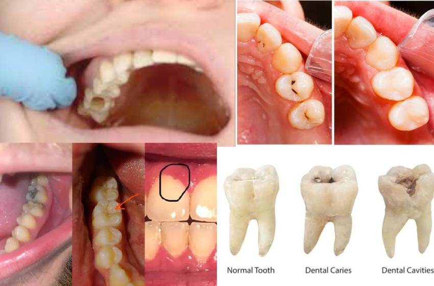
Tooth decay, cavities, or dental caries are collective problems with oral health. Most people don’t know what a cavity is, how it forms, and what stages it completes. This piece explores the visible factors of cavities, detailing how such oral health problems evolve, from when they are initially tiny white patches to the further advanced stages of infection and loss.
Table of Contents
What Are Cavities?
A cavity is a break or pit in the structure of the teeth caused by the regular weakening of the enamel by the acids produced by bacteria in the mouth. The bacteria utilize sugars from food as their source of nutrients and, in doing so, cause the production of acids that weaken and weaken the enamel. As decay progresses, it can penetrate deeper dental structures of the tooth, i.e., dentin and even pulp, causing more intricate tooth problems.
Though cavities are common in children and adults, they present differently according to their degree and stage of rot. Symptoms can be fuzzy at the start, and an awareness of the visual progression of a cavity will, at times, discourage more severe harm.
Stage 1: Enamel Demineralization and White Spots
During the early stages of decay, the first and only visible sign that a cavity is forming is when white spots appear on the tooth. They are signs of mineral loss in the enamel or demineralization. Enamel is an outer protective layer of the tooth; when bacterial acid weakens it, it loses its shine and smoothness and becomes a chalky, opaque spot.
These white dots are usually the first and most insignificant signs of tooth decay. The harm has not developed a hole or serious structure problem by this stage. To our surprise, this stage of decay can be reversed with good oral health care, application of fluoride, and changes in diet, arresting the cavity from developing further
Visual Signs:
- White, chalky patches that could look a bit dull.
- Found on the smooth surfaces of teeth or along the gumline.
- No hole is visible, but the texture could be rough or porous.
- Sensitive to acidic or sweet foods in certain instances.
Stage 2: The Brown or Black Spots Formation
The cavity becomes more evident once the enamel degenerates further and the demineralization is not reversed. The white spots become brown or black discolorations later since the decay cuts more profoundly into the enamel. The cause of the darker shade is the trapping of bacteria, food particles, and dead tissues in the decaying area.
This is the most accessible stage, even more so for the chewing sides of the rear teeth, where cavities are most apt to occur since the grooves and fissures are present. The tooth discoloration can be pale brown or darker, almost black, depending on how long the untreated decay has continued. The decay might not yet have created an observable hole, but the eroded enamel that has been done is a telling sign of forthcoming damage.
Visual Signs:
- • Brown or black discolorations that can occur in localized patches on the surface of the teeth.
- • Dark staining commonly around the edge of the teeth.
- • The tooth might feel rough, and the decay can be sensitive to touch.
- • The tooth could begin to ache upon coming in contact with cold or warm food and liquids
Stage 3: Cavities Form Observable Holes or Pits
As the tooth rot deepens, it forms a pit or hole in the tooth structure. The rot progresses past the enamel to the softer dentin layer below it. Dentin is less rot-resistant than enamel, so the cavity will progress more quickly once exposed.
At this point, the tooth will look as though it has an obvious hole or indentation, and the affected part will be covered by discolored, softened enamel. The hole is often tiny but becomes much more prominent as the decay increases. The tooth can become sensitive and tender, particularly when uncontrollable hot, cold, or sweet food or drinks.
Visual Signs:
- A visible indentation or hole in the tooth.
- Darkened or blackened coloration around the hole.
- The tooth can be soft to the touch or display a cracked surface.
- Sensitive to temperature or sweets with possible pain during chewing.
Stage 4: Deep Infection and Abscess Formation
- When the cavity is left untreated for a long time, the decay infects the tooth pulp; the innermost layer contains nerves and blood vessels. If the tooth is in this later phase, the tooth becomes vulnerable to infection, abscesses, and pus pockets that progress at the tooth root due to infection by bacteria.
- The development of the abscess will be preceded by a severe swelling of the gum covering the tooth involved and, in the most severe cases, the face or neck. The tooth will be very discolored, typically having turned black or at least dark brown, and may have cracks or fractures on its surface. During this phase, there will be sharp pains due to infection or inflammation of the pulp nerves.
- If left untreated, the infection turns foul, bringing more serious trouble such as losing the tooth or generalized to the system as a whole. The decay is no longer contained in a small portion of the tooth but has taken on a more serious and destructive form.
Visual Signs:
- Marked black discoloration of the tooth, usually covering its entire composition.
- Swelling or pus-filled abscess close to the root or gums.
- Severe pain, mostly when chewing or contacting the tooth.
- Fractures or cracks in the tooth as a consequence of its weakened condition
How Dentists Detect Cavities
Dentists employ a combination of methods to identify cavities, particularly in their early stages when they do not readily show. Perhaps the most cooperative tool is the dental X-ray, which can identify cavities between teeth or in areas that are difficult to see with the naked eye.
Through a standard check-up, a dentist will also perform a small mirror check and explore tools to examine the tooth’s outer covering for cavities or softness. An explore tool will pick up where damage from the enamel may begin without actually noticing any visible pit that is creating an opening in the tooth. Dentists also examine the overall status of the tooth and check for indications of an infection or break on the tooth’s surface.
Prevention and Treatment of Cavities
Even though cavities are a common oral disease, prevention is also possible. Good dental care, for instance, the use of fluoride toothpaste to brush teeth, flossing, and not overindulging in sugary foods, can do a great deal towards reducing the incidence of cavities. Regular dentist visits allow dentists to find decay in the early stages before becoming a serious problems.
Once a cavity has been created, the procedure consists typically of fillings, in which the decayed part of the decayed tooth is drilled away and replaced with a filling. In more extreme cases, a crown or root canal may be performed to repair the tooth. The tooth has to be removed to prevent infection if the decay spreads too far.
Conclusion
The color of a cavity changes during the development of the cavity, from the original white spot due to enamel demineralization to the later darker phases with holes and infection. The capacity to comprehend these phases and the associated color of the cavities can form a serious component of early cavity recognition, which guarantees prompt treatment before further damage occurs. A consistent routine of oral care in conjunction with expert treatment by your dentist is vital to maintaining the health and endurance of your teeth.